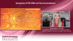
Multimodal Imaging for DR/DME
Diabetic retinal disease experts discuss the standard diagnostic imaging tools that are available to detect diabetic retinopathy and diabetic macular edema (DR/DME).
Episodes in this series

Rishi Singh, MD: We have an advent of screening tools. We've learned more about OCT [optical coherence tomography] angiography and wide field imaging using some of the cameras nowadays that have led to our insights. So what do you recommend for your standard diagnostic testing for such patients when they come in your clinic?
Carolyn Majcher, OD, FAAO: This is an area of interest for me for sure. I think incorporating multimodal imaging technologies in the assessment of patients with diabetes is really essential to meet that ever-increasing demand for diabetic eye care, as you were mentioning in the first question. This technology is going to allow us to detect the earliest pathology and vascular abnormalities so that we can make early diagnoses, and detect disease quicker and easier in their examination. It's going to allow us to see more patients in less time, but still provide the highest quality of patient care possible. I think the most important thing about multimodal imaging technology is it allows us to accurately stage the disease, but also pick up the earliest proliferative retinopathy possible. There are various multimodal imaging technologies available, including wide field retinal imaging, OCT, OCT angiography; that's primarily what I utilize, but I'm sure you also use fluorescein angiography as well.
Wide field and ultra-wide field imaging devices, whether it's color fundus photography or scanning laser ophthalmoscopy, are capable of imaging up to about 80% of the retinal surface. It's really impressive where the technology has gone. And that provides us with particular value in detecting and documenting peripheral diabetic retinopathy lesions. We know this is of some prognostic value that patients where the majority of their retinal hemorrhaging is out in the periphery are at a greater risk to progress to the proliferative stage of the disease.
OCT provides high-resolution cross-sectional images of the retinal layers and is probably most useful for detecting macular edema and has become absolutely essential in classifying macular edema as either being center-involved or non-center-involved. And then thirdly, OCT angiography provides high resolution microvascular detail that's going to highlight any sort of subtle vascular abnormalities for us that may be very subtle or perhaps invisible on our clinical fundus examination alone. These vascular abnormalities may be microaneurysms like retinal non-perfusion, or areas of small IRMA [intraretinal microvascular abnormalities]. It's also going to be useful to—especially montage OCT angiography—to visualize midperipheral nonperfusion, which if there's a substantial amount, we know there's a risk for that patient to become proliferative if they're not already because of VEGF release from that ischemic retina. But montage OCT angiography has also helped me in a lot of cases find some very subtle, small areas of neovascularization elsewhere out in the arcades that I didn't see with clinical examination alone. Because of that volumetric data set we get with OCT angiography, we can isolate out abnormal vasculature and isolate out preretinal neovascularization. So it allows us to definitively differentiate between IRMA, which is intraretinal microvascular abnormalities, a feature of perhaps moderately severe or severe non-proliferative stage vs neovascularization, which we know to be proliferative diabetic retinopathy. So I think for all those reasons, multimodal imaging is absolutely critical in the assessment of patients with diabetic retinopathy.
TRANSCRIPT EDITED FOR CLARITY
Newsletter
Don’t miss out—get Ophthalmology Times updates on the latest clinical advancements and expert interviews, straight to your inbox.


