Stages of DR/DME and Therapy Selection
Rishi Singh, MD, provides an overview of the stages of diabetic retinopathy and diabetic macular edema (DR/DME) and the retinal findings associated with each stage as well as factors to consider when selecting therapy.
Episodes in this series

Carolyn Majcher, OD, FAAO:Dr Singh, could you please describe and define the various stages of diabetic retinopathy and the retinal findings associated with each stage?
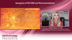
Rishi Singh, MD:We have an established classification stream going back to the diabetic retinopathy severity trials that were done in the early 1980s. This was when they established that laser was treatment necessary for patients with proliferative disease. The way I describe it to patients is the way I know it in my own practice, which is that mild and moderate diseases essentially are where the pericytes or the supportive cells in the retinal vascular are lost. You start having tight junctions, and you start developing dot and blot hemorrhages in the retina. It’s not until you get to severe nonproliferative disease where you get some of the more important findings. That’s defined as 4 quadrants of hemorrhage, 2 quadrants of venous beading, and 1 quadrant of IRMA [intraretinal microvascular abnormalities]. [Any] of those categories makes you a patient with severe nonproliferative disease.
All that is prior to the next phase, which is proliferative disease in the non–high-risk or high-risk category. I think that whole classification has gone out the window. At this point, we consider patients as having proliferative disease or not. That’s when you have neovascularization of the disc, neovascularization elsewhere, neovascularization of the iris, or even vitreous hemorrhage present in the eye. That would be a sign of proliferative disease. Each of these stages is important. The more important stages are the mild to moderate together vs the severe vs the proliferative stages, which determine progression of disease more severely. Interestingly, we find that patients with the highest risk of progression are those in the severe nonproliferative category. If they have severe nonproliferative disease, they’re highly likely to develop proliferative disease within a 1- to 2-year period of time.
Carolyn Majcher, OD, FAAO:What factors do you consider when selecting therapy for patients with diabetic retinopathy or diabetic macular edema?
Rishi Singh, MD:When we talked about this in the beginning, about the drivers for outcome, it is around baseline vision. That’s how studies have been shown to be beneficial in this category. For good vision categories, we can follow Protocol V or Protocol T where they both say in those trials done by the DRCR [Diabetic Retinopathy Clinical Research] Retina Network, that as long as the vision is very good, you can just manually observe the patient. With vision of 20/25 to 20/40, you have lots of different options for treatment, albeit bevacizumab is the cheapest option for those patients. When you get to baseline vision of 20/50 or worse, that’s when the other drugs matter. In particular, aflibercept was shown to be beneficial in the management of these patients over time, and therefore it was used for treatment for many of these patients. There is an opportunity to choose any of these drugs or at least go to one drug and then switch to another, but this is what the evidence has shown thus far from many of the randomized clinical trials we’ve been able to see.
TRANSCRIPT EDITED FOR CLARITY
Newsletter
Don’t miss out—get Ophthalmology Times updates on the latest clinical advancements and expert interviews, straight to your inbox.


