


Imaging
Latest News
Advertisement
Latest Videos


Shorts
0:43
Is there a "smoking gun" biomarker for predicting outcomes?
2 months ago
by
Michael Ip, MD(+1 more)
CME Content


Advertisement
More News

Steffen Schmitz-Valckenberg, MD, discusses how optical coherence tomography-based models may enable rapid, noninvasive assessment of functional loss in GA at Angiogenesis 2026.

China approves ZEISS ARTEVO 750 and 850 ophthalmic microscopes, expanding 3D digital visualization and iOCT-ready workflows for cataract and retinal surgery.

Andrew G. Lee, MD, and Drew Carey, MD, discuss how optic disc cupping after optic neuritis reflects nerve and ganglion cell thinning, not disease type, helping distinguish it from glaucoma.
AI reads infant retinal scans to flag lung disease early, while transient vision loss warns of looming stroke and heart events.

Optoretinography is an emerging technology used to test light-evoked photoreceptor activity.
From artificial intelligence to home monitoring, Joel Schuman, MD, of Wills Eye Hospital, explores the innovations that could change how clinicians detect and treat glaucoma in the new year.

Combining imaging and patient symptoms improves assessment of disease progression.

AAO 2025 revealed that true-color widefield imaging, AI-powered home OCT, and refined FAERS analyses are collectively transforming retinal diagnostics into a more precise, continuous, and safety-aware system.


Anat Loewenstein, MD, discusses the transformative impact of home OCT and AI on monitoring retinal diseases at AAO 2025.

Surgeons reflect on milestones that have redefined patient care—and share a glimpse of the advances that promise to shape the next era of eye health.
Deborah A. Ferrington, PhD, highlights how breakthroughs in imaging, AI, and stem cell research are reshaping ophthalmology.

The AAO 2025 meeting offered a platform for companies to showcase their latest technologies to advance ophthalmic patient care.
As ophthalmic technologies move at supersonic speed, AI and gene therapy take center stage.
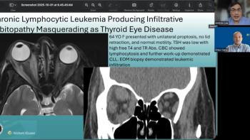
Andrew G. Lee, MD, and Drew Carey, MD, highlight how chronic lymphocytic leukemia can mimic Graves’ orbitopathy, underscoring the importance of a thorough evaluation.
Explore how digital image management and AI transform ophthalmology, enhancing diagnostics and personalizing patient care in retina practices.

Subramanian discusses how subtle retinal and optic nerve findings can point to underlying neurologic disease.
The future of diabetic retinal imaging will be impacted by the use of widefield OCT-A technology, enhancing diagnosis and monitoring while addressing current limitations.

OCT-A angiography can help reveal early neurodegenerative disease signs, offering a non-invasive method for identifying high-risk patients through retinal analysis.

Recapping the Heidelberg Engineering International SPECTRALIS Symposium—and Beyond.

Advanced imaging and awareness of systemic risk factors are essential.
Advertisement
Advertisement
Trending on Ophthalmology Times - Clinical Insights for Eye Specialists
1
DSLT for glaucoma and ocular hypertension: Real-world outcomes in 218 eyes
2
NCX 470 gains positive regulatory feedback in China ahead of US NDA submission
3
Label Updates and Dosing Flexibility in Retinal Vascular Diseases
4
First patient dosed in phase 3 pivotal cohort of LIGHTHOUSE trial evaluating ATSN-201 for X-linked retinoschisis
5