Diagnostic Tests for Dry Eye Disease
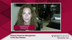
Bill B. Trattler, MD, Crystal Brimer, OD, Cynthia Matossian, MD, FACS, and Kelly K. Nichols, OD, MPH, PhD, discuss their preferred clinical diagnostic tools and tests to evaluate patients for dry eye disease (DED) and strategies for educating patients on their results.
Episodes in this series

Transcript
Bill B. Trattler, MD: So, with that in mind, I'd love to hear how everyone would diagnose dry eye, what objective tests everyone's using. So, let's go with Dr Matossian first. What tests do you feel are the most helpful for you to objectively diagnose dry eye?
Cynthia Matossian, MD, FACS: I think if you were to ask a room full of dry eye specialists what diagnostic tests they would do, each one would come up with a different kind of list. What they would do first and what they would do to follow up to see whether the treatment is effective. At Matossian Eye, what we used to do were three things. One was meibography. Everybody got meibography because to me it's like the X-ray of the lungs. When somebody shows up with a cough, I needed to see the images of the meibomian glands. You know, their health, their structure, their architecture, where they dropped out and so forth. The second one was tear osmolarity (TO), because that helped me understand kind of the surface evaporative state, how disrupted or dysregulated the tear film was. And lastly, everybody got an MMP-9 test so that I could see whether there was inflammation. According to the results, my treatment algorithm varied, and I could titrate my treatment to whether the MMP-9 was positive or negative, whether the TO was significantly abnormal or not, et cetera.
Bill B. Trattler, MD: I assume you did other tests; did you do Schirmer's test, break up time, staining? I just want to get a just a good sense of what you do besides those three diagnostic tests.
Cynthia Matossian, MD, FACS: We didn't do Schirmer's [test]. They took a long time. There was reflex tearing involved. I just, you know, I thought it was a bit too subjective. I didn't do that. But of course, [with] staining, I always do is lissamine green 100% of the time to look at the conjunctival staining, which told me a very different story than fluorescein staining on the cornea. I did both. I looked at tear meniscus height and commented on that. Of course, I looked at the lid margins and the orifices of the meibomian glands, whether they were powded or inspissated. And [I] kind of pressed on the eyelid with just a Q-Tip to see the quality and the consistency viscosity of the meibo, and of course a good slit lamp exam period.
Bill B. Trattler, MD: That's really helpful. Let's hear from Dr. Nichols and then we'll actually go through all of them, and we'll talk a little bit more. But, Dr. Nichols, tell me your approach.
Kelly K. Nichols, OD, MPH, PhD: So, I think it depends on if we're targeting somebody who hasn't done any dry eye before in terms of the recommendations, I think that what we're going to hear is what I would say is like the nirvana of a dry eye practice from our panelists here today. And so, if the goal is to try and get somebody who hasn't done any of it before, you know, the most simple approach would be some sort of assessment of symptoms, verbally usually, and then an evaluation of either staining or osmolarity or tearful breakup time. And if any one of those three is abnormal with some sort of symptomatology, which I think you can get from most anybody, then that would be a positive screening for dry eye. That was recommended by the DEWS 2 work group as the most simple, basic entry into dry eye as a screener. And then if you have the ability to do additional testing, like you have meibography, you can use that to evaluate the meibomian glands, but the simple slit lamp exam and pressing on the eyelids, of course, that is something that anybody could do in the context of their office. As well as look at the tear meniscus height as a surrogate for Schirmer because who's really going to go do a Schirmer's test in practice? So, you can kind of hit all the different areas in a simple way if you don't have that fancy equipment, which we all love, and then either refer to somebody who does, like Crystal or Cynthia or you, Bill, and so that they can have a more detailed workup. So, I think that's really two approaches for those who are just starting out. You have a great instrument in the slit lamp, and you can do a lot with it. And a thorough lit exam is really critical.
Bill B. Trattler, MD: Great comments, that's fantastic. Dr Brimer?
Crystal Brimer, OD: I completely agree. Kelly brought up a good point of sometimes we get so involved and here's the 100 things that we do because we love it, right? It is our passion. And I want to know. I can't stop myself most of the time, to be quite frank. But you make a good point. And one thing I would add to that, for somebody who doesn't have a meibographer, transilluminate, pull the lid down and look. It doesn't excuse you to not know what's going on with the glands, because in some people you can see it with just white light. If you can't, then turn this slit lamp off, put the transilluminator under that lower lid and fold it over, and you can get a decent idea of, is there truncation, is there drop out? What I would add to the list of tests that were initially listed by Dr. Matossian is, I do a cotton whisk test on every dry eye eval, and I do debridement because I want to see where any tear film debris is coming from. Is it biofilm on the lid margin? Is it poor quality meibomian that's coming out? Is it allergic mucus? What is it? And then, I use meibomian expression paddles. And I feel like that gives me the most production without a lot of discomfort at all. I go through the whole gamut of test. One that's valuable as a patient education tool is the tear film dynamic, which is basically just a video showing how much debris is in the film. That tends to get quite a response from them, and it makes them a little bit more motivated to do something. And it helps them understand why the eyes read when we have all these contaminants in there.
Bill B. Trattler, MD: Perfect. Everyone has [shared] so many great tests that can be done. I guess we put a list together. There's so many different choices that anyone doctor can do and as Dr Nichols shared, a lot of times it's something more focused. You don't need to do everything on every patient, unless you're in a totally dry center as a patient that has failed therapy elsewhere. One thing I didn't hear from anybody is pulling eyelashes for mites. Does anyone tell you eyelashes for mites? Is that important, if you see scurf and blepharitis, are you treating as if they have mites. Just curious as that's a hot topic.
Cynthia Matossian, MD, FACS: I don't pull lashes. I have the patients look down. And when you see the collarettes at the base of their lash follicle, that's almost pathognomonic for demodex blepharitis. So, I don't have to pull. I don't have a big microscope to look under to see the live mites. But looking for collarettes is really important.
Bill B. Trattler, MD: I agree. Dr. Nichols, Dr. Brimer, are you guys pulling lashes or just kind of following Dr. Matossian's plan.
Crystal Brimer, OD: I just look for the collarettes and then call it like I see it.
Kelly K. Nichols, OD, MPH, PhD Agreed.
Bill B. Trattler, MD: Perfect. That's me too. I just want to make sure I'm on the right track there. Again, just going back because I think is one of the most challenging parts, because you know, we have our listeners, our viewers, watching us and we were listing all these tests, it can be overwhelming. I'll just share that, in my typical practice, a lot of patients are coming in with specific problems [and] they're not even mentioning dry eye; your cataract evaluation patients coming in for their annual exam, their diabetic retinopathy, and they need a screening. You know, for those patients, it's much more simple. I just typically would put some fluorescein looking [at] tear breakup time and if there's corneal staining. The tear film volume, for me, is also important. Those were the two tests, beside symptoms; it's kind of what I'm doing a typical non-dry eye patient just to make sure I'm not missing dry eye that should be evaluated or should be treated. But this is really helpful because I know making the diagnosis is just part of it. Then we have to figure out how to treat these patients. Any other comments? Dr. Brimer, I know you're always excited about dry eye, did you want to make another comment? If you want to make it, go ahead.
Crystal Brimer, OD: So just like what Kelly said. All right, we've got a slit lamp, and a good slit lamp exam goes a long, long way. But if the patient can't see what you see, does it? So, you don't necessarily need the fanciest equipment out there. But I do feel like, to convert that patient to get them, to come back for an eval, or to get to ask them to do a treatment, it's a lot easier to show them a video, to show them that magnified lid margin where the collarette is there, and get a response, then it is just to talk about it. So, if you're out there and you're struggling with compliance and you're struggling with patient buy-in, I believe it's because your diagnostic tool, your way of showing them, may be lacking.
Transcript is AI-generated and edited for clarity and readability.
Newsletter
Don’t miss out—get Ophthalmology Times updates on the latest clinical advancements and expert interviews, straight to your inbox.


