Video content above is prompted by the following:
Challenges in Postrefractive Surgery Cataract Patients
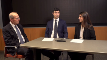
Presenter:
- Steven J. Dell, MD - Medical Director, Dell Laser Consultants, Austin, Texas
Expert Background
Steven J. Dell, MD, is an ophthalmologist and medical director of Dell Laser Consultants in Austin, Texas. This presentation was recorded at a roundtable meeting at the 2025 American Society of Cataract and Refractive Surgery Annual Meeting in Los Angeles, California.
Key Discussion Topics
- Evolving patient demographics
- Generation X patients (many with prior radial keratotomy (RK) or laser vision correction history since 1995) now entering cataract surgery age
- High patient expectations for spectacle independence
- Need for rapid assessment of patient psychology and expectations (“psychometrics”) in addition to biometrics
- Postlaser vision correction challenges
- Residual refractive error as a major cause of patient dissatisfaction
- Difficulty accurately determining corneal power in patients who have undergone postrefractive surgery
- Challenges with ocular surface disease and irregular astigmatism in patients post RK
- Enhancement dilemmas after cataract surgery
- Limitations in enhancing outcomes in patients with prior laser-assisted in situ keratomileusis (LASIK)
- Risks of lifting old LASIK flaps (epithelial ingrowth concerns first described by Andrew Caster, MD)
- Unpredictability of epithelial healing and “epithelial lensing effect” afterphotorefractive keratectomy (PRK) enhancements
Notable Insights
- Dell emphasized that “any presbyopia-correcting strategy has to have a bailout scenario” for managing suboptimal outcomes.
- Dell observed that epithelial thickness can vary dramatically (30-80 µm) across different areas of the cornea in patients who have undergone postrefractive surgery.
- The epithelium may form a “negative meniscus lens” after hyperopic laser vision correction or a “positive meniscus lens” after myopic correction, affecting refractive outcomes.
Recovery of proper epithelial thickness distribution can take 1 to 2 years after enhancement procedures, making patient management challenging.