Uveitis is uncommon, but it causes a disproportionate share of visual morbidity.

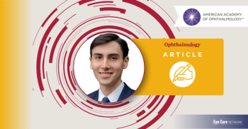
Noab Banoub, a third year medical student, describes his journey into medicine and his view of ophthalmology's future.
Noah Banoub wasn't pushed into medicine. Neither of his parents, who immigrated from Egypt, worked in health care. Medicine came to him through the tight-knit Coptic Egyptian church community where he grew up.
"A lot of my friends and their parents were actually doctors," he said. However, one stood out: the obstetrician who delivered Noah and his sister, who also gave sermons and taught Sunday school at the church. "That was probably my first introduction to medicine," he commented. Medicine was something he grew into by being surrounded by these individuals at church.
As a human biology student at the University of California, Davis, Noah volunteered at the Shifa Community Clinic, which serves underserved Middle Eastern and South Asian communities. The first real exposure to ocular disease occurred when he joined the diabetic retinopathy committee. Important lessons were the recognition of the importance and positive impact of early screening for the preventable disease and the devastating consequences when that window of opportunity slams shut.
"I didn't really connect all the dots at that time," he said. "Ophthalmology was quietly getting under my skin."
The COVID-19 pandemic compressed his college time into three years; that was followed by two gap years as a medical scribe and medical assistant in an orthopedic surgery practice. During this interlude, he learned how to talk with patients who were scared or in pain, and he watched how a surgeon reasons through problems in real time. A big recognition was the lure of imaging.
"I really loved being able to watch a scan or image change, and how that was going to change the entire plan for our patient," he said. He also valued the mixture of time in clinic and operating room, and seeing a patient's quality of life improve shortly after surgery. "I didn't really know it at the time, but I was pretty much describing ophthalmology before I even considered it."
During those same years, his grandfather was diagnosed with age-related macular degeneration (AMD). Because his grandfather's English is limited, Noah accompanied him to appointments and served as a translator. He explained what the injections were for and how often they would be needed. That experience helped him see how much ophthalmology had in common with the orthopedic work he enjoyed.
Enrollment in the Kaiser Permanente Bernard J. Tyson School of Medicine in Pasadena, CA, in the school's fifth entering class was a boon because of the small class sizes. "Because of that, I could explore different specialties early and not be funneled down a predetermined path."
Research was a medical school requirement, and a mandatory scholarly project led him to his mentor. His chosen project was progression of macular telangiectasia type 2 (MacTel). "Admittedly, I didn’t know what it was, but it sounded like an interesting topic," he said. The project's mentor was Robin Vora, MD, a retina specialist at Kaiser Permanente Oakland.
"It was probably one of the best things that happened to me in medical school," Noah said. "Dr. Vora is so great in terms of how much he invests in students, in his clinic, and in his patients."
Noah was involved in a study designed to track how MacTel progresses across Kaiser Permanente Northern California. The team identified about 750 patients with a MacTel diagnosis and planned to use optical coherence tomography (OCT), fundus imaging, and visual acuity to see whether injections or comorbidities such as hypertension and diabetes changed the course of the disease.
As attending physicians reviewed the cases, a pattern emerged. "They were realizing, oh, this patient doesn't actually have MacTel; this patient also doesn't have MacTel," Noah said. On review, about 192 of the roughly 750 patients had been misdiagnosed.
The misdiagnoses came from an outdated view of the disease. MacTel was long thought of as a vascular disorder, but it is now understood mainly as a neurodegenerative disease of the Müller cells. As a result, clinicians who saw microaneurysms, hemorrhages, cystoid changes, or leakage sometimes jumped to MacTel. Most of the misclassified cases turned out to be neovascular AMD, retinal vein occlusion, or hypertension-related microaneurysms, he explained.
"Not everything that has microaneurysms is MacTel; not everything that has cystic changes is MacTel," he said. "It really requires a longitudinal look at the imaging, and different types of imaging, to make that diagnosis."
In addition, no single biomarker establishes the diagnosis, he added, and diagnosis depends on following findings over several years of OCT imaging, including changes in the ellipsoid zone and the angle and branching pattern of the retinal vessels. "We're trying to get ophthalmologists to realize that the minute they see something vascular, they shouldn't automatically think MacTel," he said. The team has presented the findings as an abstract at several meetings, and a manuscript is under review.
Getting the diagnosis right became more urgent with the FDA approval of revakinagene taroretcel (Encelto, Neurotech Pharmaceuticals, Inc.) in March 2025, the first approved treatment for MacTel type 2. The treatment is a surgically implanted, encapsulated cell therapy that slowly releases ciliary neurotrophic factor, a neuroprotective agent. Noah's team began seeing patients receive the implant in August 2025.
Because the therapy is costly and requires invasive surgery, the team wants multimodal imaging used to confirm eligibility. "Otherwise, we're giving patients with the wrong diagnosis a surgery that's invasive," he said. The goals are better outcomes, fewer misdiagnoses, and shorter diagnostic delays. He also wants clinicians to know clearly when to refer patients to a retina specialist.
"For years, MacTel type 2 was a diagnosis of exclusion and no approved therapy was available," he said. "Now that treatment exists, getting the diagnosis right isn't just an academic exercise anymore. It's really what determines whether a patient is eligible for something that can preserve their vision."
Noah's interest in health equity goes back to his time at Shifa. He is now helping launch a study at Kaiser Permanente Northern California on persistent inflammation after cataract surgery in high-risk patients.
After surgery, patients can choose either a steroid injection or a tapering course of steroid drops, the latter of which is preferred. Patients who already had postoperative iritis or uveitis often don't trust another injection. The study will try to determine at why some patients fail the steroid taper.
Published studies have suggested that African-American, Asian-American, and Hispanic patients are at higher risk. The team hopes to identify high-risk patients before surgery and counsel them differently. If the chart review supports it, that could mean giving an injection right away instead of waiting for a taper to fail. Earlier work suggests that approach resolves inflammation faster.
Right now, the choice of injection or taper and when to start and stop, is determined by each surgeon. "If we can prove that works better than the taper, we want to see if we can standardize that protocol across all of Kaiser's cataract surgeries," Noah said.
Noah commented on both how rapidly MacTel type 2 went from being a diagnosis of exclusion without a therapy to recognition of its frequent misdiagnosis despite imaging, which is exactly the gap their research was focusing on: showing how often MacTel is misdiagnosed on imaging and how quickly a treatment was approved and is being implanted in eligible patients.
He used this example to describe ophthalmology as a whole.“I think this is the path down which ophthalmology is headed: earlier, more precise diagnosis through multimodal imaging, and AI-assisted analysis of this imaging is on the rise. Pairing that with disease-modifying therapy instead of just symptom management seems to be where we're headed.”
He continued, “On the flip side, my work with quality improvement and health equity has shown me that the future of the field isn't just about new technology; it's about making sure the existing, effective care we have reaches the patients who need it, regardless of their background,” he emphasized.